

Thin Facial Bone
The bone covering the front surface of an upper incisor root can be extremely thin. Preserving this bone is important because it helps support the gum contour around the future implant crown.
Replace a missing or failing tooth with a digitally planned dental implant designed to restore natural appearance and function. Treatment is planned with the final ceramic restoration in mind from the beginning.
One dental implant + one prefabricated abutment + one all-ceramic crown
For eligible single-tooth replacement cases.
Replacing a front tooth requires careful coordination of surgery, esthetics, and function. Implant position, available bone, gum contours, tooth shape, shade, translucency, bite, and the patient's smile are considered together before treatment begins.
The bone covering the front surface of an upper incisor root can be extremely thin. Preserving this bone is important because it helps support the gum contour around the future implant crown.
Front teeth often have an angled relationship between the visible crown and the natural root. Implant position therefore has to be planned around both the available bone and the intended position of the final ceramic tooth.
The height and shape of the gum around a front tooth strongly affect how natural the final result appears. Preserving the tissues from the beginning can be especially important in patients with a visible smile line.
The final restoration must reproduce tooth shape, surface texture, shade, translucency, and proportions while also fitting comfortably within the patient's bite.
When the anatomy and condition of the extraction site allow it, Dr. Andrews may remove the damaged tooth and place the dental implant during the same visit rather than waiting several months before a second implant surgery.
Combining extraction and implant placement can shorten the overall treatment process and reduce the number of separate surgical procedures. For the patient, that can mean fewer surgical appointments, fewer recovery periods, and less time spent moving through repeated phases of treatment and healing.
Immediate placement is not appropriate for every site. The decision depends on available bone, the condition of the socket, infection, implant stability, gum anatomy, and the planned final restoration.
In selected cases, Dr. Andrews may combine immediate implant placement with specialized techniques intended to preserve the thin facial bone and soft-tissue contours that help determine the final appearance of a front tooth.
With Partial Extraction Therapy, sometimes called the socket-shield technique, a carefully prepared thin portion of the natural root is intentionally retained on the facial side of the socket.
That small root segment keeps its attachment to the surrounding tissues and may help preserve the thin bone and gum contour that would otherwise tend to shrink after the tooth is removed.
Some post-extraction bone remodeling occurs along the outside surface of the socket. In selected cases, Dr. Andrews may place a thin protective barrier along the outer surface of the facial bone.
The technique is designed to reduce this external bone-remodeling process during healing and help maintain the width and contour of the ridge supporting the implant restoration.
The objective is to preserve as much of the patient's existing anatomy as possible while replacing the failing tooth. Depending on the individual case, immediate implant placement may be combined with Partial Extraction Therapy, periosteal inhibition, bone grafting, or other tissue-management techniques.
By planning these steps together, treatment may require fewer separate surgical procedures while maintaining the bone and gum architecture needed for a natural-looking final restoration.
A clinical examination and 3D evaluation are used to determine whether immediate implant placement or tissue-preserving techniques are appropriate for your individual anatomy.
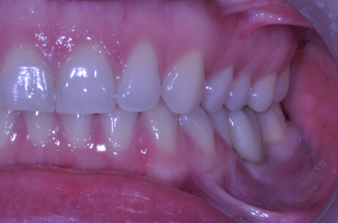
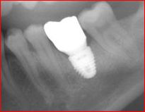
The following case demonstrates treatment of a broken upper front tooth using immediate implant placement and coordinated management of the surrounding bone, soft tissue, and final ceramic restoration.

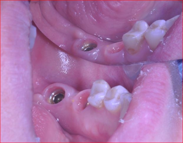
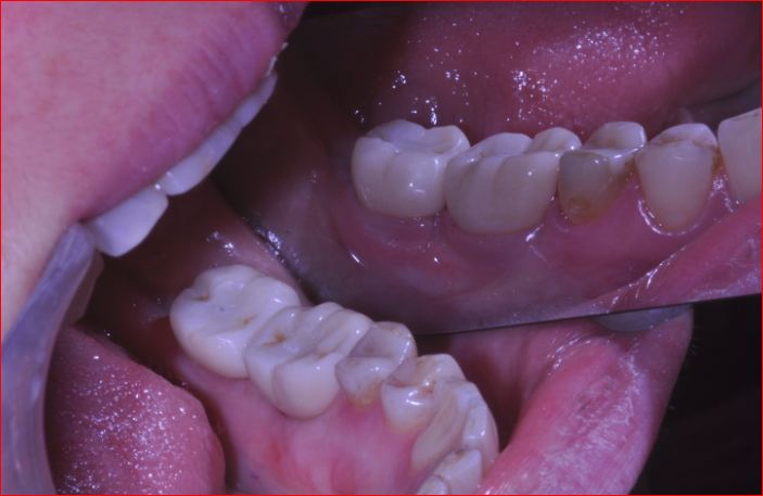
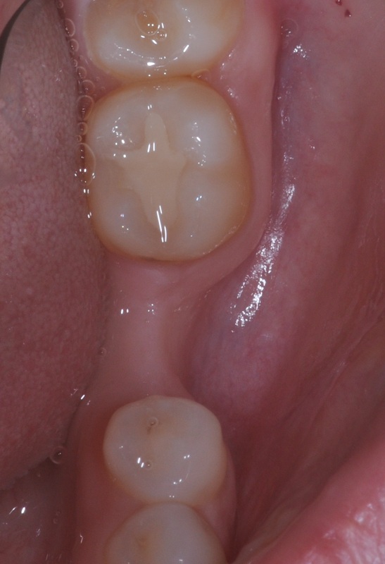
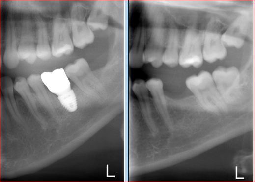
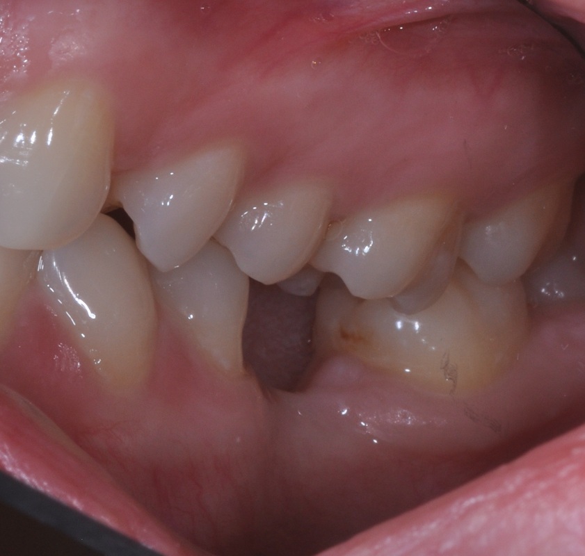
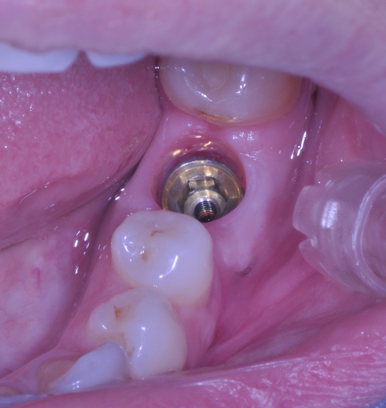
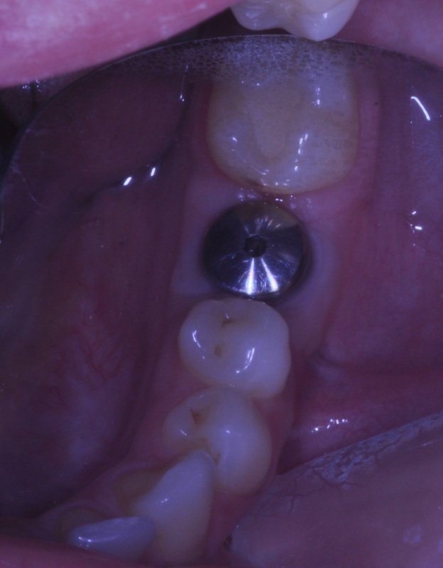
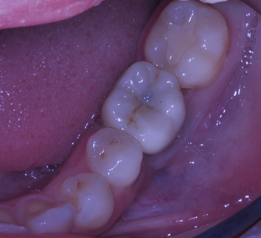
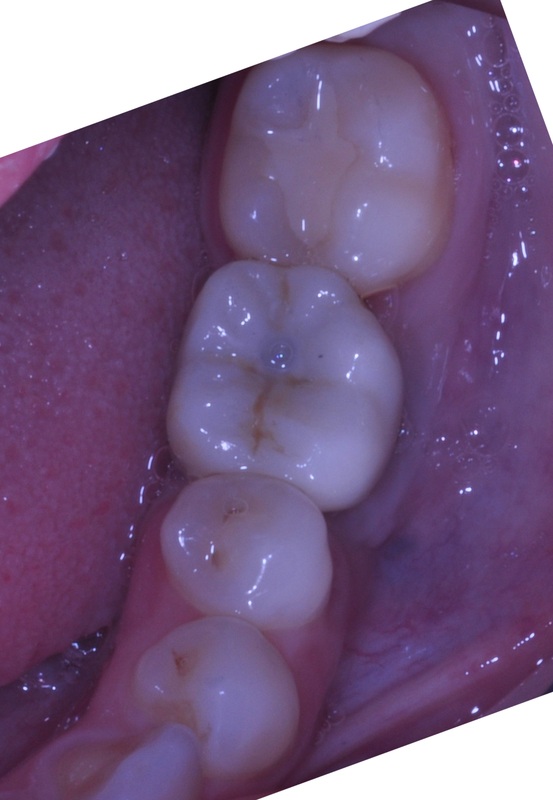
Replacing a missing molar or premolar with a dental implant begins with evaluating the tooth, surrounding bone, neighboring teeth, and the final restoration. Whenever possible, implant treatment should be considered before a compromised tooth is removed.
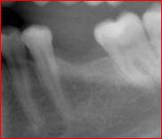
Natural tooth roots help maintain the width of the surrounding bone. After extraction, significant bone-volume loss can occur, particularly in the horizontal dimension.
Planning ahead may help preserve more treatment options for future implant placement.
Adequate bone is required to support a dental implant. When the ridge is too narrow or deficient, bone grafting may be required before or at the time of implant placement.
Nearby teeth and supporting tissues should also be healthy before treatment proceeds.
Under favorable conditions, complete implant treatment including the final crown may take approximately five months in the lower jaw and six months in the upper jaw.
Treatment may take substantially longer when additional bone reconstruction or staged procedures are necessary.
In many implant cases, Dr. Andrews is able to coordinate tooth extraction, bone grafting when necessary, and implant placement during the same surgical visit. Whether this is appropriate depends on the condition of the tooth, available bone, infection status, anatomy, and the individual treatment plan.